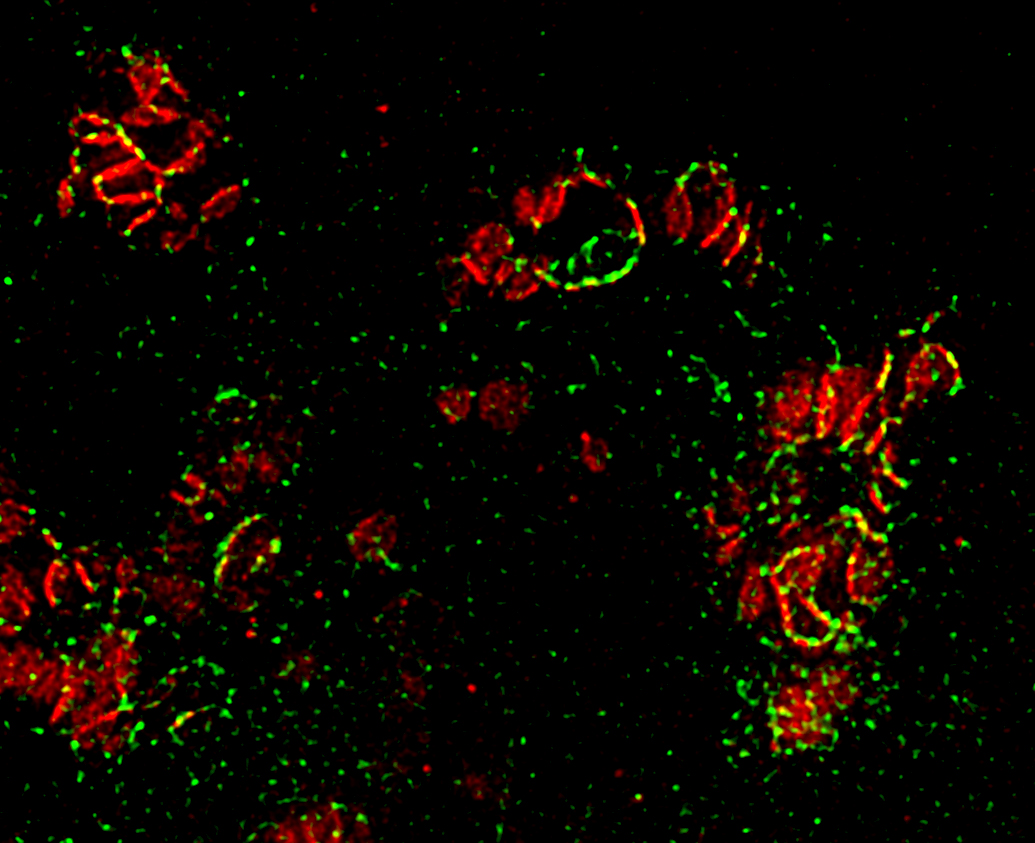
Confocal STED microscopy image of parkin (green) and p62 (red) decorating mitochondria in HeLa cells following depolarisation with Carbonylcyanide-3-chlorophenylhydrazone (CCCP).
Fluorescence microscopy image showing TOM20-positive mitochondrial-derived vesicles (red), indicated by arrowheads, budding off from the mitochondrial network in the neuroblastoma SH-SY5Y cell line.